3D Cone Beam Technology
This new technology has raised expectations in both dental care and dental treatment. 3D imaging provides more accurate diagnostics and treatment precision, resulting in better patient understanding and satisfaction.
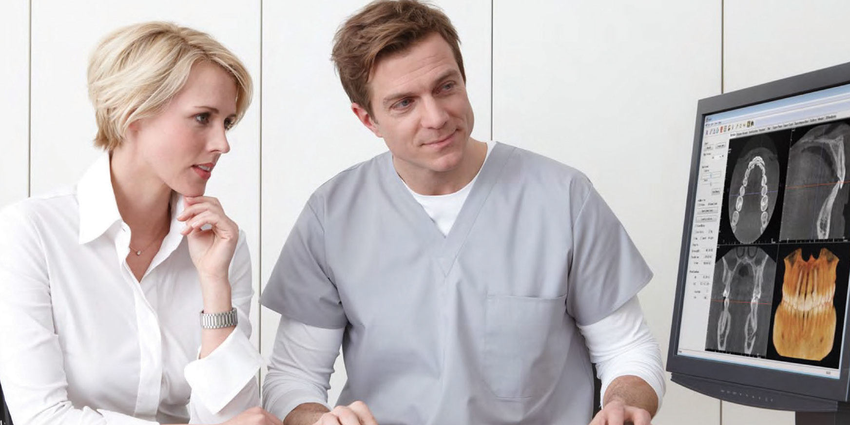
3D dental imaging can be fast and simple. After a quick scan, we are able to view the patient’s dental anatomy instantly. This advancement in dentistry allows us to gather accurate and complete information in order to treat patients more effectively.
At The Nottingham ConeBeam Centre, we are equipped with the latest GENDEX GX-DP 700 cone beam dental CT scanner. CT scans can provide the clinician with 3D visualisation of the anatomy of the jaws and associated structures, to improve planning for procedures.
What is the Difference Between 3D and Regular Dental X-rays?
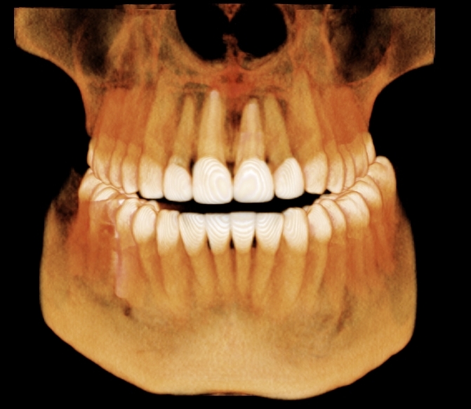
A 3D x-ray creates a volume of data allowing us to ‘slice’ through the anatomy at any angle, revealing anatomy and issues that are sometimes hidden when using 2D imaging. A 3D dental imaging system can give us a more complete view of the dental anatomy at nearly every angle.

Benefits of 3D X-rays
The use of a 3D imaging machine give us the confidence to know we are diagnosing and treating issues correctly. Plus, we can install confidence in our patients, too, by educating them using 3D x-rays.
A 3D dental imaging system can give us the most detailed information possible for more accurate diagnosis and treatment planning. The more information we have, the better care we are able to provide our patients. The process is quick, simple and completely painless, giving us a comprehensive view of the targeted dental anatomy with just one 10-20 second scan.
The move to digital radiography and cone beam 3D imaging goes well beyond just saving time and has opened many doors for us to expand our practice and the services we are able to provide to your patients.
- Targeted area can be viewed from a wide variety of angles
- Faster, easier and more precise images
- Anatomy can be ‘sliced’ to view at the optimal angle
- Significant time savings for everyone in the practice, including the patient
- Top-quality reading and scanning capabilities, resulting in better treatment planning
- Unlimited views of the targeted areas in one pass
- More complete picture of oral structures, including jaw bones, soft tissues and nerve paths, which allows us to develop treatment plans with more precision and accuracy
- Provides dentists the ability to operate on impacted teeth and plan for implant placements
Benefits for the patient
There are several advantages for a patient undergoing a 3d X-ray examination. The major ones are less exposure to radiation compared to medical CT scans and time saved. Only one scan is needed to get a complete image of the targeted dental anatomy. In most cases, this all happens in less than one minute. Plus, we are able to provide diagnosis and treatment courses in the span of a single appointment.
Patients may not even realize they are receiving another major benefit: Education. 3D imaging forms a virtual model of the patient’s anatomy by utilizing computer-automated rendering software to translate the scan data into something almost everyone can understand. Now we can show the patient the condition of their teeth, soft tissues and jaws right on a computer screen. We can also pinpoint problem areas by turning and rotating the model, then show the patient what their facial structure the final outcome once the treatment is complete.
